
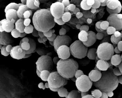
Electron microscopy is an expensive and laborious, but powerful technique to study nanoparticles. In electron microscopy the electron beam is focussed on the sample and scanned over a defined area of the sample. Images are constructed based on electrons coming from the sample surface. Number-weighted size distributions of nanoparticles are analyzed by identifying the nanoparticles individually. In order to have a representative particle size distribution, quite some nanoparticles should be investigated. The added value of electron micrsocopy for nanoparticle assessment is that besides the particle size of the nanoparticles also the shape of the nanoparticles can be visualized.
The nanoparticle shape helps in a further quantification and interpretation of the specific surface area results obtained by BET surface area analysis. The shape of the nanoparticles namely determines the threshold value of the volume specific surface area obtained for the BET surface area by gas adsorption. Although BET surface area analysis is considered as a screening method, electron microscopy has proven to be a confirmatory method following European Commission’s guidelines on nanoparticles identification. This is due to the fact that the BET surface area is a indicator value from which potential nanoparticle presence is derived, whereas the actual presence of nanoparticles can directly be seen in electron microscopy and the “seeing is believing” statement is applicable.
In contrast to the differential centrifugal method and dynamic light scattering for nanoparticle analysis, the sample needs to be dry in electron microscopy analysis similar to the volume specific surface area assessment. The sample volume used in electron microscopy analyses is very limited and therefore requires a critical representative sampling step prior to the actual electron microscopic investigation.We have dedicated sampling equipment for representative sampling in our laboratory. Fortunately, nanoparticles are very small and thus one easily encounters a large number of particles even in a very small sample volume.
Electron Microscopy Analysis FAQs
Electron microscopy is used to directly visualise nanoparticles and determine their size, shape and morphology. By analysing individual particles in electron microscope images, a number-based particle size distribution can be created.
Scanning electron microscopy, or SEM, is primarily used to examine the surface structure and morphology of particles. Transmission electron microscopy, or TEM, uses electrons transmitted through a thin sample and can provide detailed information about small particles and their internal structure. The most suitable technique depends on the material and the information required.
Yes, electron microscopy can be used to measure the size of individual nanoparticles from images. It can also show the particle shape and morphology. To obtain a representative particle size distribution, a sufficient number of particles must be analysed from a properly selected sample.
Electron microscopy analysis generally requires the sample to be prepared in a dry form. This differs from techniques such as DLS and DCS, which analyse particles dispersed in a liquid. Sample preparation must be carefully controlled to avoid altering the particle structure or causing artificial agglomeration.
Only a very small amount of material is examined during electron microscopy. If the selected portion is not representative of the entire sample, the resulting particle size distribution may be misleading. Careful sampling and analysis of a sufficiently large number of nanoparticles are therefore essential.


